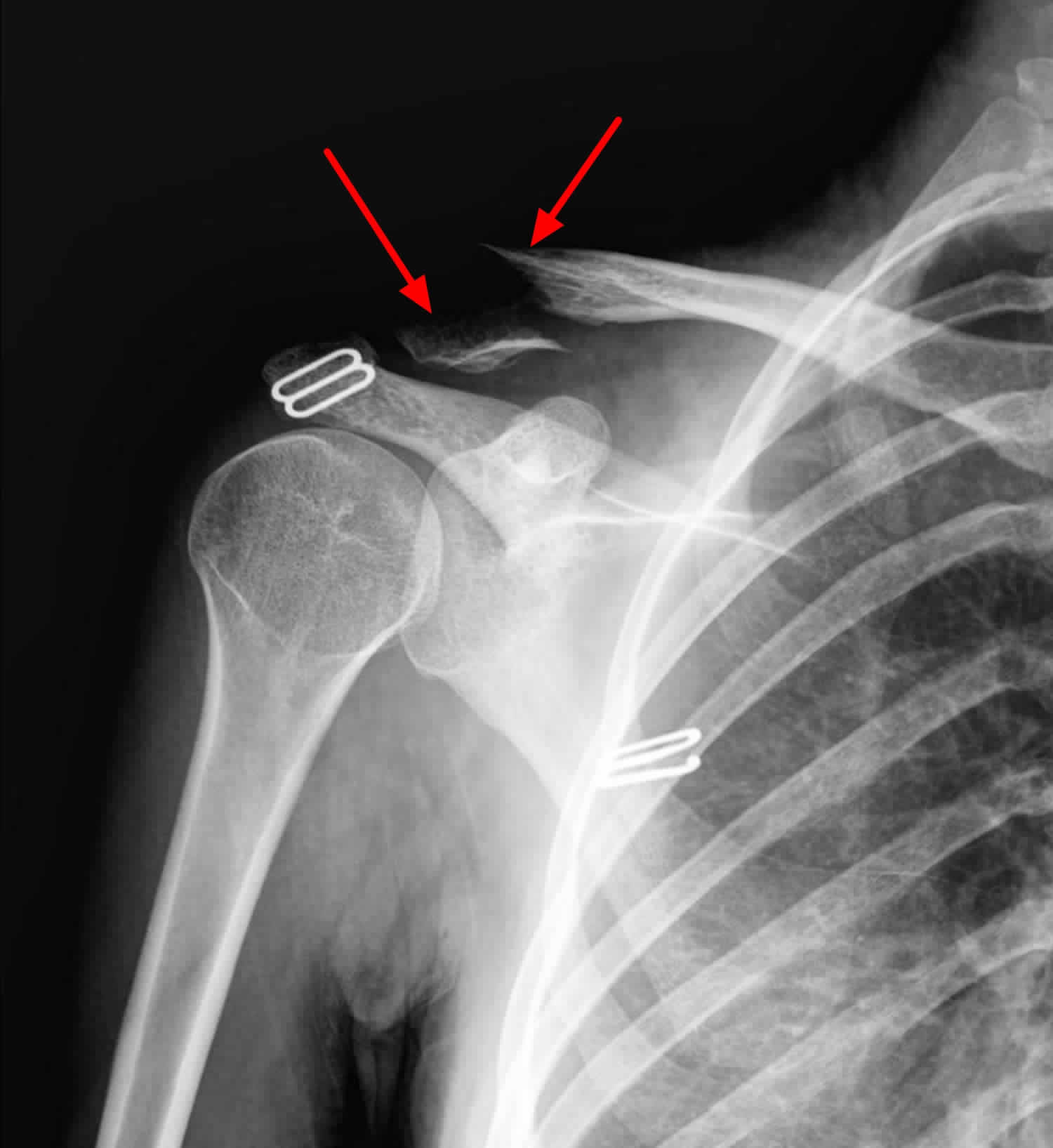
Collarbone X Ray . the clavicle (collarbone) is one of the most fractured bones in the body. To learn more about the. The ap clavicle is often indicated in patients with suspected clavicular injuries following trauma such as falling onto ones side. To help pinpoint the location of the fracture; The posterior shoulder should be in contact with image receptor (ir) or tabletop, without rotation of body. The standard ap view of the clavicle is taken with the patient upright or sitting, with arms at the sides, chin raised, and looking straight ahead. imaging of the clavicle. Symptoms of a broken collarbone include severe pain and swelling at the site of the fracture and with visible deformity in some cases. It can be requested as part of a. the radiographic series of the clavicle is utilized in emergency departments to assess the clavicle, acromioclavicular and.
from healthjade.net
the clavicle (collarbone) is one of the most fractured bones in the body. To learn more about the. The ap clavicle is often indicated in patients with suspected clavicular injuries following trauma such as falling onto ones side. To help pinpoint the location of the fracture; Symptoms of a broken collarbone include severe pain and swelling at the site of the fracture and with visible deformity in some cases. The posterior shoulder should be in contact with image receptor (ir) or tabletop, without rotation of body. It can be requested as part of a. imaging of the clavicle. The standard ap view of the clavicle is taken with the patient upright or sitting, with arms at the sides, chin raised, and looking straight ahead. the radiographic series of the clavicle is utilized in emergency departments to assess the clavicle, acromioclavicular and.
Broken collarbone or clavicle fracture signs, symptoms and treatment
Collarbone X Ray the radiographic series of the clavicle is utilized in emergency departments to assess the clavicle, acromioclavicular and. To help pinpoint the location of the fracture; The posterior shoulder should be in contact with image receptor (ir) or tabletop, without rotation of body. The standard ap view of the clavicle is taken with the patient upright or sitting, with arms at the sides, chin raised, and looking straight ahead. The ap clavicle is often indicated in patients with suspected clavicular injuries following trauma such as falling onto ones side. Symptoms of a broken collarbone include severe pain and swelling at the site of the fracture and with visible deformity in some cases. To learn more about the. the radiographic series of the clavicle is utilized in emergency departments to assess the clavicle, acromioclavicular and. imaging of the clavicle. the clavicle (collarbone) is one of the most fractured bones in the body. It can be requested as part of a.
From www.drdavidduckworth.com.au
Clavicle Fractures Dr David Duckworth Collarbone X Ray The posterior shoulder should be in contact with image receptor (ir) or tabletop, without rotation of body. To learn more about the. The ap clavicle is often indicated in patients with suspected clavicular injuries following trauma such as falling onto ones side. imaging of the clavicle. the radiographic series of the clavicle is utilized in emergency departments to. Collarbone X Ray.
From santabarbarasportsorthopedic.com
Clavicle Fracture Surgery Shoulder Surgeon Santa Barbara, Santa Collarbone X Ray the clavicle (collarbone) is one of the most fractured bones in the body. It can be requested as part of a. Symptoms of a broken collarbone include severe pain and swelling at the site of the fracture and with visible deformity in some cases. The posterior shoulder should be in contact with image receptor (ir) or tabletop, without rotation. Collarbone X Ray.
From www.alamy.com
Collarbone x ray hires stock photography and images Alamy Collarbone X Ray Symptoms of a broken collarbone include severe pain and swelling at the site of the fracture and with visible deformity in some cases. The ap clavicle is often indicated in patients with suspected clavicular injuries following trauma such as falling onto ones side. To learn more about the. To help pinpoint the location of the fracture; the clavicle (collarbone). Collarbone X Ray.
From pressbooks.pub
Clavicle Fracture Undergraduate Diagnostic Imaging Fundamentals Collarbone X Ray The ap clavicle is often indicated in patients with suspected clavicular injuries following trauma such as falling onto ones side. the clavicle (collarbone) is one of the most fractured bones in the body. The posterior shoulder should be in contact with image receptor (ir) or tabletop, without rotation of body. The standard ap view of the clavicle is taken. Collarbone X Ray.
From www.dreamstime.com
Xray of the Right Collarbone. Fracture of Clavicle. Stock Photo Collarbone X Ray imaging of the clavicle. To learn more about the. The posterior shoulder should be in contact with image receptor (ir) or tabletop, without rotation of body. The standard ap view of the clavicle is taken with the patient upright or sitting, with arms at the sides, chin raised, and looking straight ahead. It can be requested as part of. Collarbone X Ray.
From www.sciencephoto.com
Fractured collar bone, Xray Stock Image M330/1356 Science Photo Collarbone X Ray To help pinpoint the location of the fracture; the radiographic series of the clavicle is utilized in emergency departments to assess the clavicle, acromioclavicular and. It can be requested as part of a. imaging of the clavicle. To learn more about the. Symptoms of a broken collarbone include severe pain and swelling at the site of the fracture. Collarbone X Ray.
From www.dreamstime.com
Shoulder Xray, Clavicle (collarbone) Fracture Stock Image Image of Collarbone X Ray the radiographic series of the clavicle is utilized in emergency departments to assess the clavicle, acromioclavicular and. The posterior shoulder should be in contact with image receptor (ir) or tabletop, without rotation of body. To help pinpoint the location of the fracture; imaging of the clavicle. The ap clavicle is often indicated in patients with suspected clavicular injuries. Collarbone X Ray.
From www.alamy.com
Fractured collarbone, Xray Stock Photo Alamy Collarbone X Ray To help pinpoint the location of the fracture; The posterior shoulder should be in contact with image receptor (ir) or tabletop, without rotation of body. the radiographic series of the clavicle is utilized in emergency departments to assess the clavicle, acromioclavicular and. The standard ap view of the clavicle is taken with the patient upright or sitting, with arms. Collarbone X Ray.
From fracturehealing.ca
Clavicle Fractures Fracture Healing Collarbone X Ray imaging of the clavicle. The standard ap view of the clavicle is taken with the patient upright or sitting, with arms at the sides, chin raised, and looking straight ahead. The posterior shoulder should be in contact with image receptor (ir) or tabletop, without rotation of body. To learn more about the. The ap clavicle is often indicated in. Collarbone X Ray.
From www.alamy.com
FRACTURED COLLARBONE, XRAY Stock Photo Alamy Collarbone X Ray the radiographic series of the clavicle is utilized in emergency departments to assess the clavicle, acromioclavicular and. To help pinpoint the location of the fracture; The ap clavicle is often indicated in patients with suspected clavicular injuries following trauma such as falling onto ones side. the clavicle (collarbone) is one of the most fractured bones in the body.. Collarbone X Ray.
From www.gettyimages.com
Fractured Collarbone Xray HighRes Stock Photo Getty Images Collarbone X Ray the clavicle (collarbone) is one of the most fractured bones in the body. The ap clavicle is often indicated in patients with suspected clavicular injuries following trauma such as falling onto ones side. To learn more about the. Symptoms of a broken collarbone include severe pain and swelling at the site of the fracture and with visible deformity in. Collarbone X Ray.
From www.alamy.com
Fractured collar bone, Xray Stock Photo Alamy Collarbone X Ray the clavicle (collarbone) is one of the most fractured bones in the body. The ap clavicle is often indicated in patients with suspected clavicular injuries following trauma such as falling onto ones side. It can be requested as part of a. To learn more about the. Symptoms of a broken collarbone include severe pain and swelling at the site. Collarbone X Ray.
From www.what0-18.nhs.uk
Clavicle (Collar Bone) Fracture Healthier Together Collarbone X Ray the radiographic series of the clavicle is utilized in emergency departments to assess the clavicle, acromioclavicular and. To learn more about the. It can be requested as part of a. The standard ap view of the clavicle is taken with the patient upright or sitting, with arms at the sides, chin raised, and looking straight ahead. Symptoms of a. Collarbone X Ray.
From www.dreamstime.com
Xray of the Left Collarbone. Fracture of Clavicle. Consolidation of Collarbone X Ray It can be requested as part of a. The ap clavicle is often indicated in patients with suspected clavicular injuries following trauma such as falling onto ones side. the radiographic series of the clavicle is utilized in emergency departments to assess the clavicle, acromioclavicular and. To help pinpoint the location of the fracture; imaging of the clavicle. . Collarbone X Ray.
From www.alamy.com
Fractured collar bone, Xray Stock Photo Alamy Collarbone X Ray To learn more about the. It can be requested as part of a. the radiographic series of the clavicle is utilized in emergency departments to assess the clavicle, acromioclavicular and. the clavicle (collarbone) is one of the most fractured bones in the body. The standard ap view of the clavicle is taken with the patient upright or sitting,. Collarbone X Ray.
From healthjade.net
Broken collarbone or clavicle fracture signs, symptoms and treatment Collarbone X Ray The posterior shoulder should be in contact with image receptor (ir) or tabletop, without rotation of body. the radiographic series of the clavicle is utilized in emergency departments to assess the clavicle, acromioclavicular and. the clavicle (collarbone) is one of the most fractured bones in the body. To learn more about the. The standard ap view of the. Collarbone X Ray.
From radiopaedia.org
Clavicle fracture Image Collarbone X Ray The ap clavicle is often indicated in patients with suspected clavicular injuries following trauma such as falling onto ones side. the clavicle (collarbone) is one of the most fractured bones in the body. The standard ap view of the clavicle is taken with the patient upright or sitting, with arms at the sides, chin raised, and looking straight ahead.. Collarbone X Ray.
From www.sciencephoto.com
Broken collar bone, Xray Stock Image M330/1017 Science Photo Library Collarbone X Ray It can be requested as part of a. the radiographic series of the clavicle is utilized in emergency departments to assess the clavicle, acromioclavicular and. Symptoms of a broken collarbone include severe pain and swelling at the site of the fracture and with visible deformity in some cases. To learn more about the. The posterior shoulder should be in. Collarbone X Ray.
